The Process of How “Light” Becomes “Sight”
 The eye’s design allows it to focus, capture, and convert information from light into signals that can be delivered to the brain and interpreted, or “seen,” as an image. The eye works much like a production line in this sense, with the end product being an image in your brain.
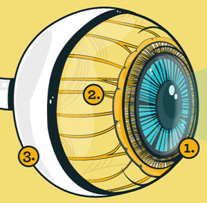
1. When light first hits the eye, it must pass through the thin layer of tears that covers the cornea. It enters the cornea or surface layer of the eye. Like a clear window, the cornea focuses the light and allows it to penetrate the eye.
Directly between the cornea and pupil is another layer of moisture. This thicker layer of moisture is called the aqueous humor. This layer of moisture circulates through the eye, keeping a constant pressure and giving the eye its spherical shape.
Once the light is past this layer, it passes through an aperture-like structure called the pupil. This dark circle in the middle of the iris may contract or dilate to limit or increase the amount of light that passes deeper into the eye.
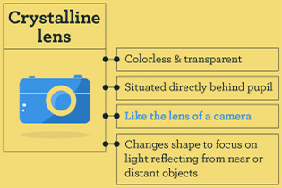
 Situated directly behind the pupil is colorless, transparent crystalline lens. Like the lens of a camera, this structure changes shape to focus on light reflecting from near or distant objects. Ciliary muscles surround the lens and hold it in place. When these muscles relax, they flatten the lens, allowing the eye to see objects that are far away. The ciliary muscle contracts in order to thicken the lens and see closer objects clearly.
2. The focused beam of light now enters the center of the eye. The light is once again plunged into moisture called vitreous humor. The retina surrounds this clear jellylike substance and contains the photoreceptors that are the light’s final destination.
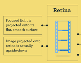
 The retina is the inner lining of the back of the eye. Like a movie screen or the film of a camera, the focused light is projected onto its flat, smooth surface. Interestingly, the image projected onto the retina is actually upside down and the brain flips it 180 degrees as it’s interpreting the image to form the correct picture.
3. There are millions of light-sensitive photoreceptors in an eye, and they come in two main varieties: rods and cones.
Rods are useful for monochrome vision in poor light, whereas cones are used to detect color and fine detail. Cones are packed into a part of the retina known as the fovea. Like a bulls-eye on a dartboard, the fovea is located at the dead center of the retina and contains a full 10% of the specialized light sensitive nerve endings necessary for reading and detail work.
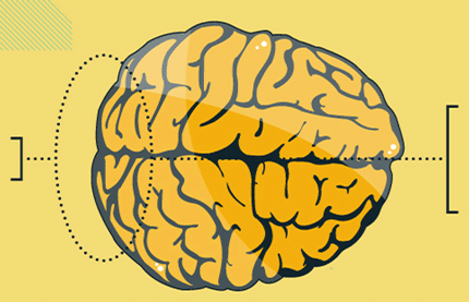
When light strikes the rods or cones in the retina, it’s immediately converted into an electro-chemical signal that is instantly relayed to the vision center near the back of the brain via the optic nerve. The brain then translates these signals into the images we see. That’s how the eye works, and it all happens faster than the blink of an eye.
